Light-induced chloroplast movements in leaves:
seeing is believing
INTRODUCTION
Light-induced changes in the cellular distribution or orientation
of chloroplast's have been observed in nearly all green plants including algae,
mosses, ferns, and angiosperms. Under low light conditions, chloroplasts accumulate
along the cell walls that are perpendicular to the incident light. Under high
light conditions, they accumulate along the walls that are parallel to the incident
light. These are the regions of plant leaf cells where internal fluence rates
of light are the highest and lowest, respectively, and it is believed likely
that the light-induced chloroplast movements serve an adaptive function. In
algae, moss and ferns, both red and blue light can cause chloroplast movements.
In terrestrial angiosperms, chloroplast movements have been shown to be blue-light-specific
but the red/far-red phytochrome photoreceptors appear to play a role in modulating
the response. In any event, chloroplast movements can have visible affects on
light transmittance and reflectance properties of leaves. For example, Wada
and Sugai (1994) exposed fern gametophytes masked with stenciled letters to
high fluence rates of light and produced visible images of the letters in the
fern gametophytes. In a more recent study in which the blue light photoreceptor
phot2 was described, Kagawa et al. (2001) showed that images could also be observed
in Arabidopsis leaves. The light-induced chloroplast movements are sensitive
and robust enough that by using masks made from black and white photographs,
it is possible to obtain detailed images in living leaves. In the case of chloroplast
movement images, the grain size is that of the individual chloroplasts rather
than the starch granules contained within them. Moreover, chloroplast movement-induced
images can be observed in living leaves without the need for chemical stains
and the same leaf can be used over and over to create new images simply by changing
the masking image and re-exposing the leaf to light.
The procedure described to create images in leaves by chloroplast
movements provides a simple project that can be done in classrooms or laboratories.
The procedure produces a striking visible demonstration of an important adaptive
response in plants and it clearly illustrates how plants can rapidly respond
to changes in their environment.
THE PROCEDURE
1. First you will need to find a suitable leaf to work with. Coleus
leaves can be excellent as long as you can find a variety that doesn't have
too much purple pigmentation (the anthocyanains absorb the blue light required
to cause chloroplasts to move and can make seeing the effect difficult). Geranium
leaves also work but they are quite think and it is more challenging to see
the images. However, the thickness adds an interesting 3-D effect. Although
they are pretty small, Arabidopsis leaves work well.
2. Once you have a source of leaves, you need to prepare some images to use
as templates. For this, you need black & white images with high contrast.
To start, it is recommended that you use text until you have established that
you leaves and lighting system will work. Once you have confirmed that it is
working, print some high-contrast photos or clip-art onto transparency film
with a standard black & white laser printer (do not use ink-jet printers
since the ink will wash off the film).
3. Cut a leaf from the plant and place it on wet paper towel. If you have Petri
dishes, place the wet paper towel on the inside of the lid, place the leaf on
the paper, then place the laser printed transparency on the leaf. Cover the
leaf and transparency with the inverted bottom of the Petri dish as shown in
the images below. If you don’t have Petri dishes, wrap the wet paper towel,
leaf and transparency in plastic wrap. It is important to keep the leaf wet
during the next step.
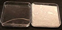
A. Petri dish lid with wet paper towel
|
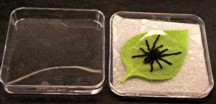
B. Petri dish lid with wet paper towel and laser-printed transparency
on coleus leaf
|
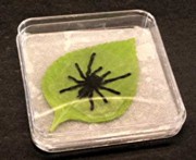
C. Same as B with base of Petri dish inserted in lid.
|
Note, you can use clear plastic wrap instead of a
Petri dish to keep the leaf from drying out during the process. |
4. Shine a bright light source on the leaf. A slide projector works great for
this. To use a slide projector, stand the tape the top and bottom of the Petri
dish together and stand it on its edge about 2 feet from the projector with
the leaf surface facing the lens of the slide projector. Turn on the light and
illuminate the leaf for about 45 min.
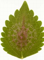
5. Remove the leaf from the wrapping and view the leaf to see the image created
by light-induced chloroplast movements. Where the light was bright, the leaf
should be more transparent than were the leaf was covered by the dark areas
of the image as shown in the example below (the NSF logo was printed with clear
lettering on a black background).
6. To facilitate viewing of the chloroplast-created image in leaves, try using
backlighting. A slide viewer works great but simply holding the leaf several
inches above some white paper can often work. Another way to make the image
stand out a little more is to look at the leaf through a blue filter or with
blue backlighting. Because chlorophyll absorbs blue light strongly, the blue
filter can increase the contrast making it easier to see the image. A digital
camera can be used to record the leaf image.
7. If the leaf is kept moist and placed in darkness or left fully exposed to
a uniform light, the image will fade as the chloroplasts move to similar positions
in all of the cells. The same leaf can be used multiple times to record new
images, even without waiting for the chloroplasts to move to erase the previous
image.
|
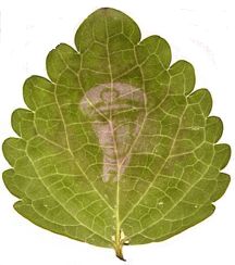
It is even possible to recreate photographs in a living leaf as shown
in the image to the right.
Created from a family photograph (Andrew J. Hangarter) |
 |
SELECTED REFERENCES
Gorton HL, Williams WE, Vogelmann TC (1999) Chloroplast movement
in Alocasia macrorrhiza. Physiol Plant 106: 421-428
Haupt W, Scheuerlein R (1990) Chloroplast movement. Plant Cell
Environ 13: 595-614
Inoue Y and Shibata K (1973) Light-induced chloroplast rearrangements
and their action spectra as measured by absorption spectroscopy. Planta 114:
341-358
Kagawa T, Sakai T, Suetsugu N, Oikawa K, Ishiguro S, Kato T, Tabata
S, Okada K, and Wada M (2001) Arabidopsis NPL1: A Phototropin Homolog Controlling
the Chloroplast High-Light Avoidance Response. Science 291: 2138-2141
Wada M and Sugai M (1994) Photobiology of ferns. In: Kendrick
RE, Kronenberg, GHM (eds). Photomorphogenesis in Plants, pp 783-802. Kluwer
Academic Publishers, Netherlands